
The visual system is a complex and highly specialised sensory system that enables organisms to perceive and interpret their environment through light. It involves intricate anatomical structures, neural pathways, and physiological processes that work in harmony to convert light into meaningful visual experiences. This post explores the anatomy of the eye and visual pathways, the role of photoreceptors (rods and cones), and the mechanisms underlying colour vision, including the opponent-process theory.
Anatomy of the Eye and Visual Pathways
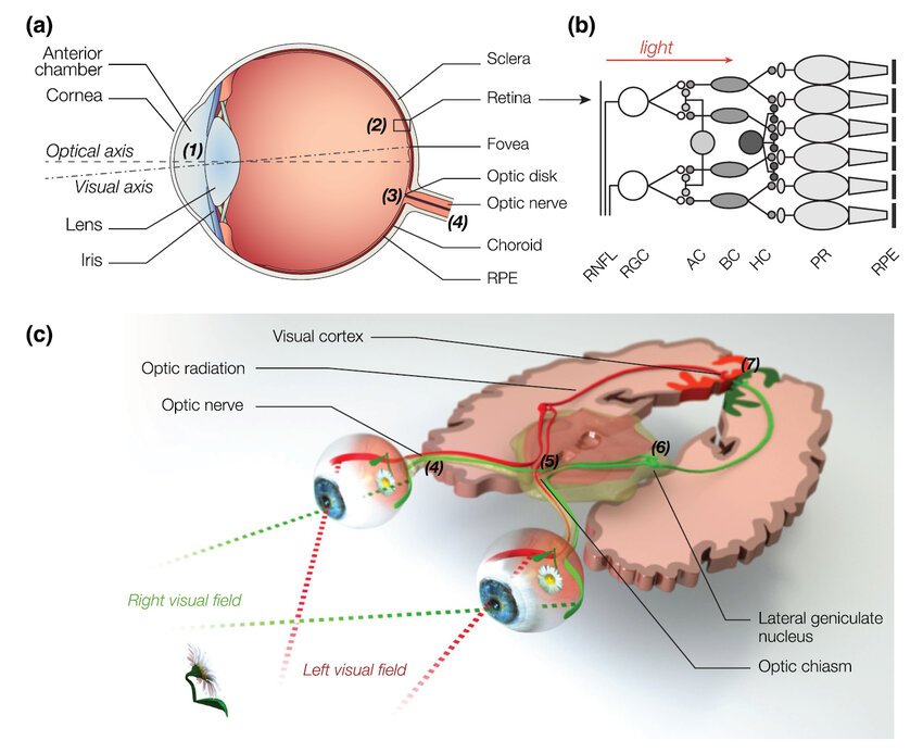
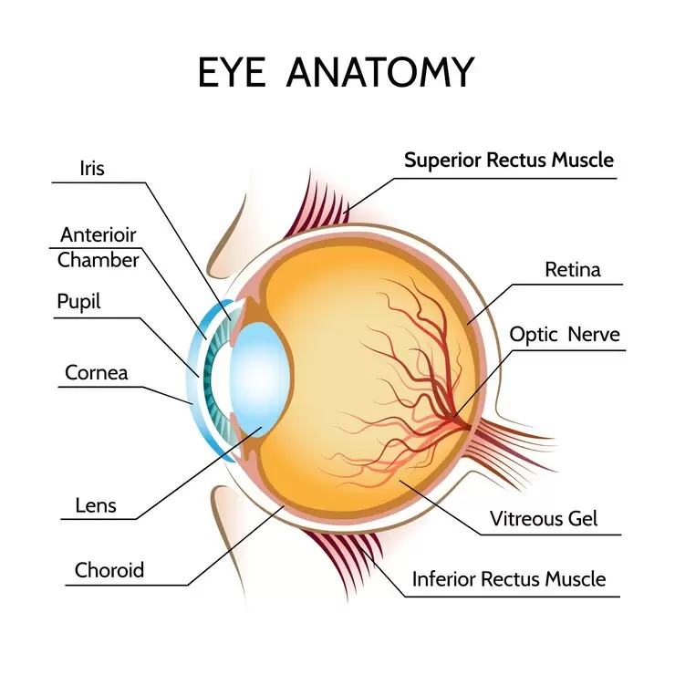
Structure of the Eye
The human eye is a remarkable organ in the visual system designed to capture, focus, and process light. Its primary components include:
- Cornea: The transparent outer layer that refracts light entering the eye.
- Iris: The colored part of the eye that controls the size of the pupil, regulating the amount of light that enters.
- Pupil: The opening in the center of the iris that adjusts in response to light intensity.
- Lens: A flexible, transparent structure that fine-tunes the focus of light onto the retina.
- Retina: The light-sensitive layer at the back of the eye, containing photoreceptor cells (rods and cones) and other neurons.
- Optic Nerve: Transmits visual information from the retina to the brain.
Visual Pathways
The visual pathway refers to the neural route by which visual information is transmitted from the retina to the brain. The process involves several key stages:
- Retinal Processing: Photoreceptors in the retina convert light into electrical signals, which are then processed by bipolar cells and ganglion cells.
- Optic Chiasm: The point where the optic nerves from each eye partially cross, allowing the brain to receive input from both eyes.
- Lateral Geniculate Nucleus (LGN): A relay center in the thalamus that processes visual information before sending it to the primary visual cortex.
- Primary Visual Cortex (V1): Located in the occipital lobe, this region is responsible for initial visual processing, such as detecting edges, motion, and orientation.
The integration of these pathways ensures that visual stimuli are accurately interpreted, enabling depth perception, object recognition, and spatial awareness.
Photoreceptors in the Human Visual System: Rods and Cones
The human retina contains two primary types of photoreceptor cells: rods and cones. These specialised cells are responsible for converting light into neural signals, enabling vision under varying conditions of illumination. Each type of photoreceptor has distinct structural and functional characteristics, tailored to specific aspects of visual system.
Rods
Rods are photoreceptor cells that are exquisitely adapted for low-light vision, a capability referred to as scotopic vision. These cells are indispensable for nocturnal and dim-light environments, where their high sensitivity to light allows for the detection of minimal illumination. The following features delineate the unique properties of rods:
- High Sensitivity: Rods are remarkably sensitive to light, capable of detecting even a single photon. This extraordinary sensitivity is facilitated by the photopigment rhodopsin, which undergoes a conformational change upon absorbing light, initiating the visual transduction process. This attribute makes rods indispensable for night vision.
- Distribution: Rods are predominantly located in the peripheral regions of the retina, with their density decreasing towards the central fovea. This distribution enhances peripheral vision, allowing for the detection of motion and objects in the periphery, even in low-light conditions.
- Monochromatic Vision: Unlike cones, rods do not contribute to colour vision. They contain only one type of photopigment, rhodopsin, which is sensitive to a broad spectrum of light but does not discriminate between wavelengths. Consequently, rod-mediated vision is achromatic, rendering the world in shades of grey.
Cones
Cones are the photoreceptor cells responsible for colour vision (photopic vision) and high visual acuity. They are optimally functional under bright-light conditions and are concentrated in the central retina, particularly the fovea. The salient characteristics of cones include:
- Colour Discrimination: Cones are equipped with three distinct types of photopigments, or opsins, each sensitive to different wavelengths of light: short (blue), medium (green), and long (red). This trichromatic system enables the perception of a wide spectrum of colours by combining signals from these three cone types.
- High Acuity: The fovea, a small depression in the centre of the retina, is densely packed with cones and devoid of rods. This anatomical arrangement ensures that light is focused directly onto cones, facilitating sharp, detailed vision. The high spatial resolution of cones is critical for tasks requiring precision, such as reading or recognising faces.
- Light Requirements: Cones operate most effectively in well-lit environments. Their sensitivity to light is significantly lower than that of rods, rendering them less useful in dim conditions. However, their ability to function in bright light ensures vibrant colour perception and fine detail discrimination during daylight.
Phototransduction
The process by which rods and cones convert light into neural signals is termed phototransduction. This intricate biochemical cascade involves several sequential steps:
- Light Absorption: The photopigments in rods (rhodopsin) and cones (opsins) absorb photons of light. This absorption triggers a conformational change in the photopigment, activating it.
- Biochemical Cascade: The activated photopigment initiates a signal transduction pathway, leading to the closure of sodium ion channels in the photoreceptor cell membrane. This results in hyperpolarisation of the cell, a reduction in the release of neurotransmitters such as glutamate.
- Signal Relay: The hyperpolarisation signal is transmitted to bipolar cells, which in turn communicate with ganglion cells. The axons of ganglion cells converge to form the optic nerve, which carries the visual information to the brain for further processing and interpretation.
In summary, rods and cones are specialised photoreceptors that collectively enable vision across a broad range of lighting conditions. Rods excel in low-light environments, providing sensitive, monochromatic vision, while cones dominate in bright light, delivering high-acuity, colour-rich perception. The phototransduction process unites these cells in their shared function of converting light into the neural language of the brain, thereby underpinning the remarkable faculty of sight.
Colour Vision and Opponent-Process Theory
Trichromatic Theory
The trichromatic theory, proposed by Young and Helmholtz, suggests that color vision is mediated by three types of cones (S, M, and L cones). The brain interprets color based on the relative activation of these cones. For example:
- Equal activation of all three cones produces the perception of white.
- Selective activation of L and M cones results in yellow or red hues.
Opponent-Process Theory
Ewald Hering’s opponent-process theory complements the trichromatic theory by explaining how colour perception is processed at the neural level. It posits that colour vision is governed by opposing pairs of colours:
- Red-Green: Neurons are excited by red and inhibited by green (or vice versa).
- Blue-Yellow: Neurons are excited by blue and inhibited by yellow (or vice versa).
- Black-White: This pair accounts for brightness perception.
This theory explains phenomena such as afterimages and colour blindness, where the balance between opponent colours is disrupted.
Integration of Theories
Modern research integrates both theories:
- The trichromatic theory explains color detection at the photoreceptor level.
- The opponent-process theory describes how signals from cones are processed in the retina and brain to produce color perception.
Conclusion
The visual system is a marvel of biological engineering, combining precise anatomical structures with sophisticated neural processing. From the initial capture of light by the eye to the intricate pathways that relay information to the brain, each component plays a critical role in shaping our visual experiences. The interplay between rods and cones allows for both night vision and vibrant colour perception, while theories like trichromacy and opponent-process provide frameworks for understanding how we perceive the world in colour. By studying these mechanisms, we gain deeper insights into the complexities of human vision and the potential for addressing visual impairments.
Discover more from Decroly Education Centre
Subscribe to get the latest posts sent to your email.